This website uses cookies so that we can provide you with the best user experience possible. Cookie information is stored in your browser and performs functions such as recognising you when you return to our website and helping our team to understand which sections of the website you find most interesting and useful.
Category:
Artificial Intelligence
Industry:
Laboratory Diagnostics
Client
The client is an innovative medtech company focused on leveraging artificial intelligence in gastrointestinal imaging diagnostics. The company primarily develops tools to enhance capsule endoscopy (CE) analysis—a minimally invasive method for imaging the small intestine that generates between 50,000 and 70,000 images per examination. They provide solutions to clinics and hospitals facing growing demand for fast and accurate diagnoses amid limited staffing resources.
With increasing clinical demand and expanding applications of this method, there is an urgent need for intelligent automation that shortens analysis time, enhances diagnostic accuracy, and ensures process scalability.
Challenge
The company identified several critical barriers limiting the efficiency and diagnostic utility of CE examinations:
- Prolonged image review times, leading to clinician fatigue and workflow bottlenecks.
- Diagnostic inconsistencies due to human subjectivity and lack of standardized interpretation.
- Missed or delayed identification of subtle yet clinically significant abnormalities.
- Inefficient analysis of large image datasets, most of which do not contain diagnostic information.
- Low representation of rare pathological findings in training data, affecting model performance in detecting such cases.
To address these challenges, the company developed an AI-driven solution that combines real-world data with synthetic data augmentation to ensure accurate classification and streamline workflows.
Solution
The core AI technology is based on advanced convolutional neural networks (CNNs) and image analysis algorithms specifically designed for the capsule endoscopy (CE) workflow. Key features include:
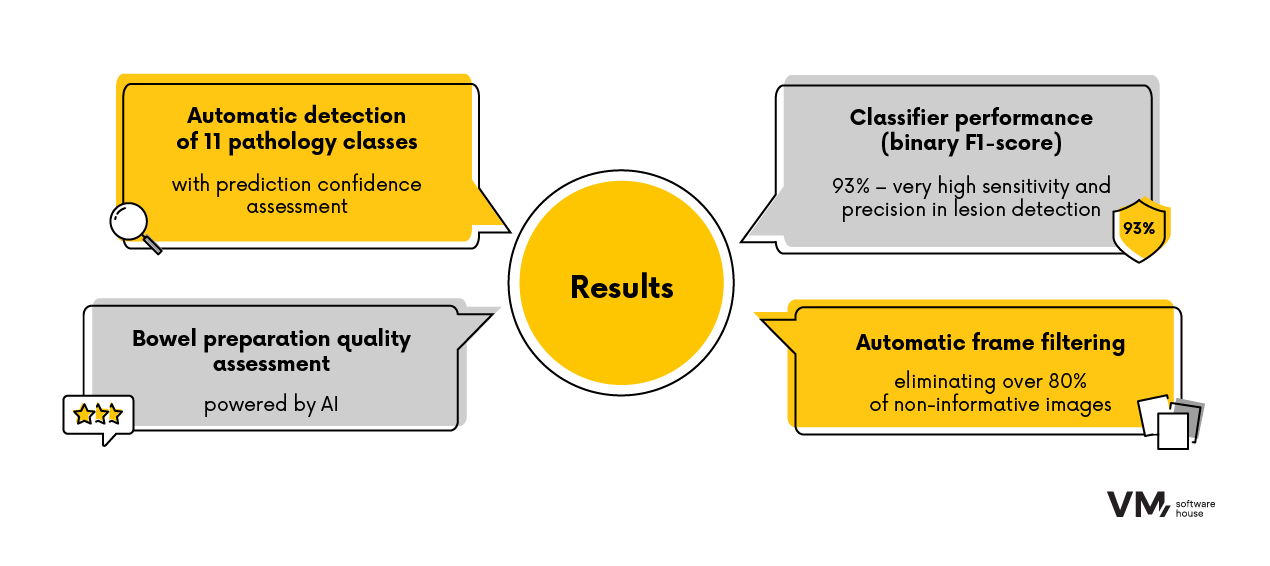
- Automatic detection of 11 pathology classes, with confidence scoring for each prediction.
- AI-based bowel cleanliness assessment, ensuring consistency in image quality evaluation.
- Automatic frame filtering, eliminating over 80% of non-informative images while retaining over 90% of frames containing pathological findings.
- High classifier performance (binary F1-score of 93%), indicating strong sensitivity and precision in detecting abnormalities.
To further enhance classification accuracy—especially for rare pathologies—the company employs a dual synthetic data generation strategy:
- Classic image transformations: Includes morphological adjustments, controlled noise, brightness normalization, and artificial lesion overlays based on real clinical images.
- Generative models (GANs, Diffusion models): Trained to produce realistic pathology images across all 11 classes, significantly improving data diversity.
This hybrid approach increases the model’s generalizability and boosts its effectiveness in identifying rare conditions.
The system also incorporates explainable AI (XAI) methods such as GradCAM, which highlight image regions crucial for model decisions, allowing clinicians to validate prediction credibility.
Model Architecture and Training
The model uses unconventional techniques to maximize sensitivity and precision in the challenging medical domain. Initial training employed self-supervised learning techniques tailored to the unique nature of CE imagery, which differs significantly from standard visual data.
Collaboration with medical experts enabled the incorporation of domain-specific knowledge, including group-equivariance (rotation invariance), which enhances performance in detecting rare pathological patterns.
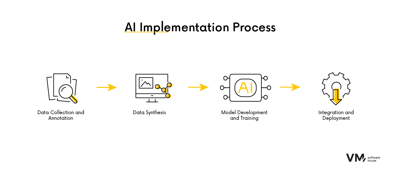
Deployment Process
Data Collection and Annotation
- Over 5 million CE images collected.
- More than 300,000 images annotated by clinical experts.
- Standardized pathology labels based on consensus guidelines.
Data Synthesis
- Traditional augmentation techniques applied to minority classes.
- Physician-validated synthetic images generated via GANs.
- Balanced training dataset combining real and synthetic images.
Model Development and Training
- Optimized CNN architecture designed for CE-specific challenges.
- Regular performance evaluation using F1-score, Precision, Recall, AUROC, and confusion matrix analysis.
Integration and Implementation
- AI engine integrated into the proprietary CE analysis platform via modular APIs.
- System includes interactive image previews and confidence metrics for classification.
Results
The AI system delivers significant clinical and operational benefits:
- 80% of images automatically filtered, drastically reducing review time.
- 90% of pathological images retained post-filtering, ensuring high diagnostic reliability (clinicians maintain full access—no pathology is hidden).
By applying advanced AI algorithms and innovative synthetic data generation techniques, the company has revolutionized capsule endoscopy diagnostics, achieving faster, more accurate, and scalable assessments. This approach not only boosts clinician efficiency but also enhances patient care by enabling earlier and more consistent detection of gastrointestinal abnormalities. The project was delivered by specialists from BioCam, a company specializing in the application of artificial intelligence in gastrointestinal imaging diagnostics.

Automating Workshop Management with an AI Assistant Based on a Large Language Model (LLM)
Design, Development, DevOps or Cloud – which team do you need to speed up work on your projects?
Chat with your consultation partners to see if we are a good match.